Technology
Position MRI™ (pMRI™)
- Weight-bearing studies of the human anatomy.
- Unrestricted range-of-motion for flexion, extension, rotation and lateral bending.
- Brain scans with the patient upright (vertical)
- Patient positioning plays a critical role in detecting clinically significant pathology.
- Multi-Position MRI Benefits
- Scans patients in flexion, extension, rotation and lateral bending
- Scans patients with the weight of the body on the spine and other supporting joints
- Scans patients in sitting positions
- Scans patients lying down
- Scans cardiovascular patients upright in their position of symptoms
- Scan patients with cerebrovascular insufficiency in the upright position of symptoms
- An unobstructed view from inside the magnet. There is nothing in front of the patient's face.
- 0.6 Tesla field strength
- Iron-frame electromagnet
- Front-open and top-open magnet design
- Magnetic poles are on the left and right of the patient
- Horizontal (transaxial) magnetic field orientation
- 18 inch (46 cm) pole-to-pole, horizontal gap
- Multi-positional capabilities for positional MRI™ (pMRI™) studies
- Translation function advances the upright or recumbent patient into center of the magnet
- Elevator function places standing patient's anatomy of interest at magnet isocenter
- Tilt function rotates the patient from upright to the recumbent position
- Table slightly tilted at 5 degrees to stabilize patients during upright scanning
- Support fixtures for RF receiver coils integrated into the table.
- Motorized and computer-controlled
- Fully MRI-compatible
- The only true Open MRI
- The most non-claustrophobic whole-body Open MRI.
- Walk in, stand or sit for the scan, walk out
- Removable seat
- An unprecedented degree of patient comfort because there is an unobstructed view of the scanner room from inside the magnet. There is nothing in front of the patient's face.
- Movable, trans-polar stabilization bars to enhance patient comfort and stability during multi-positional scans such as flexion, extension, rotation and lateral bending.
- Whisper Gradients™ for quiet scans
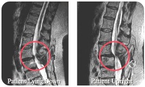
Here are two scans of a patient with low back pain. The patient had surgery but the pain and symptoms continued to get worse. The image on the left was made with the patient lying down. It shows a normal alignment of the vertebrae. The image on the right, which was done Upright, revealed that the patient had a dramatic spinal instability that the lying–down scan did not reveal and that the first surgery did not address. Following visualization of the spinal dislocation seen by the Open Upright MRI, the patient underwent surgery a second time. The patient has been pain free ever since.